
Medicine is built on understanding. But understanding looks different depending on who is asking the question.
Education Space turns real clinical information into a shared visual environment that can be explored in clinical depth, taught in context, or explained with clarity.
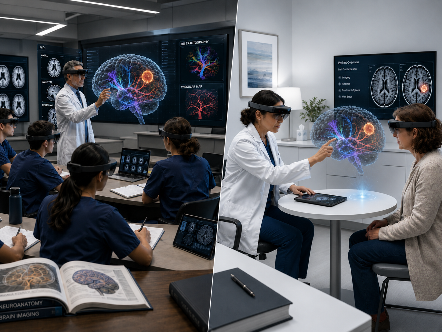
Neurospace Education Space preserves the clinical source while adapting how information is explored and communicated. The goal is not to simplify medicine into abstraction, but to make the same underlying case useful to the people who need to understand it.
Explore anatomy, pathology, functional relationships, and procedural context with the depth expected in a clinical environment. Patient-specific imaging remains central to the learning experience.
Clinical DepthMove from textbook anatomy to case-based understanding. Faculty can guide learners through real imaging, spatial relationships, annotations, and the reasoning behind clinical decisions.
Guided LearningUse patient-specific visualizations to explain what is happening, where it is happening, and how a planned procedure relates to the patient’s own anatomy—supporting clearer clinical conversations and informed-consent discussions.
Personal UnderstandingEducation Space is designed around multimodal clinical information. MRI, CT, PET, fMRI, DTI, and related clinical data can be brought into a common visual context so anatomy, function, pathology, and connectivity can be explored together.
The same case can then be presented with different levels of detail without disconnecting the learner or patient from the underlying clinical source.
In a clinical curriculum, a scan is more useful when learners can understand the structures, pathways, pathology, symptoms, and procedural consequences surrounding it.
Education Space can support faculty-led case reviews, anatomy instruction, neuroscience education, procedural demonstrations, and interdisciplinary teaching using a common spatial workspace.
Clinical imaging is information-rich, but it is rarely presented in a form designed for the person whose anatomy it represents. Education Space creates a visual layer for physician-patient communication without disconnecting the explanation from the real case.
Clinicians can use patient-specific 3D views to orient the patient, identify the relevant anatomy or pathology, and explain how a planned procedure or treatment relates spatially to surrounding structures.
A diagnosis or treatment discussion can be difficult to process when a patient is trying to understand unfamiliar anatomy, medical terminology, risks, and options all at once. By grounding the conversation in the patient’s own imaging and a clinician-led visual explanation, Education Space can help reduce uncertainty and anxiety while giving the patient a clearer basis for discussing treatment choices with their care team.
Patient education can reduce anxiety by replacing uncertainty with a clearer understanding of what is happening, what the physician is seeing, and what to expect next. Patient-specific visualizations make unfamiliar anatomy and treatment concepts more tangible, helping patients feel more oriented, prepared, and confident during the treatment conversation.
Visual explanations can show where a treatment acts, what surrounding structures matter, and why different approaches may carry different considerations.
Greater clarity can help patients ask better questions, understand benefits, risks, and alternatives, and participate more confidently in shared treatment decisions.
Education Space extends the Neurospace collaboration philosophy into teaching. A faculty member, trainee, specialist, or remote participant can share a common visual context instead of relying only on slides, screen-sharing, or disconnected screenshots.
The result is a teaching environment organized around the case itself—where the discussion remains linked to anatomy, imaging, and clinical context.
Interactive 3D anatomy can be examined at scale, from multiple viewpoints, with clinically relevant layers revealed as needed.
AR / MR VISUALIZATIONMultiple users can conceptually occupy the same visual case, enabling guided instruction, discussion, and collaborative annotation around patient-specific anatomy.
MULTI-USERSpatial experiences should remain restrained and clinically readable, with headset-based visualization used where appropriate rather than decorative holograms visible to everyone in the room.
HOLOLENS 2 / META QUEST 3 WHERE APPROPRIATEThe Neurospace approach is designed around interoperability with the imaging and clinical infrastructure institutions already use. Education Space can draw from the same ecosystem of medical imaging, case data, and collaboration tools rather than forcing education into a disconnected content silo.
Select a perspective to see how the same patient-specific case can be organized for a different educational purpose.
Explore multimodal imaging, anatomy, pathology, functional relationships, and procedural context with the level of detail required for clinical discussion and advanced training.
From advanced clinical teaching to patient communication, Education Space turns the same medical information into a visual environment designed for the person who needs to understand it.