
MAP FUNCTION.
PLAN WITH
FORESIGHT.
Functional neurosurgery operates where anatomy, circuitry, symptoms, and millimeters converge. Neurospace brings those relationships into one patient-specific environment for deep brain stimulation, epilepsy and sEEG workflows, functional mapping, stereotactic targeting, and neuromodulation—combining multimodal imaging, functional data, pathway modeling, artificial intelligence, spatial visualization, and connected planning so the surgical team can understand not only where a target is, but what surrounds it, what a trajectory may influence, and how a network may behave.
THE TARGET IS ANATOMICAL.
THE OUTCOME IS NETWORKED.
In functional neurosurgery, a coordinate is only the beginning. A DBS target must be understood in relation to circuitry and trajectory. An epilepsy hypothesis may require sEEG electrodes to sample distributed regions and reveal seizure onset and propagation. The clinical question therefore extends into circuits, white-matter pathways, functional territories, symptom networks, electrode geometry, trajectories, and structures that must be protected. Neurospace is designed to make those relationships visible together, in the individual patient rather than as disconnected reference images.
SEE THE PATIENT
Bring structural MRI, CT, diffusion imaging, tractography and other available patient data into one registered planning context.
SEE THE NETWORK
Relate candidate targets to structural connectivity and functionally meaningful pathways instead of treating deep anatomy as isolated points.
SEE THE CONSEQUENCE
Compare target and trajectory options against proximity, functional anatomy and modeled pathway relationships before finalizing a plan.
KEEP THE WORKFLOW CONNECTED
Carry planning context into multidisciplinary review, neuronavigation and collaborative workflows without rebuilding the case from scratch.
ONE PATIENT.
MULTIPLE SIGNALS. ONE CLINICAL CONTEXT.
Neurospace aligns the data that functional neurosurgery depends on and presents it as a coherent patient-specific model. The objective is not to replace specialist interpretation, but to reduce the cognitive friction of mentally reconciling separate datasets.
REGISTERED DATA LAYERS
Anatomical reference for deep nuclei, ventricular boundaries and surrounding structures.
Available hardware, frame or localization context incorporated into the same coordinate environment.
White-matter pathways displayed relative to target candidates and trajectories.
fMRI, TMS or other available functional information can be aligned with anatomical planning context.
Symptoms, prior procedures, notes and case-specific priorities can inform how relevant data are presented to the team.
ARTIFICIAL INSIGHT
FOR FUNCTIONAL DECISIONS.
MULTIMODAL SYNTHESIS
Align and organize structural imaging, diffusion data, functional datasets and available clinical context into a patient-specific planning model.
FUNCTIONAL–ANATOMICAL CORRELATION
Surface spatial relationships between candidate targets, deep anatomy, functional territories and structural connectivity for specialist review.
TARGET INTELLIGENCE
Compare patient-specific target candidates against configured anatomical, pathway and case parameters without presenting the result as an autonomous clinical decision.
PATHWAY + TRAJECTORY ANALYSIS
Model how proposed trajectories relate to critical structures and selected tracts, helping the team examine alternative approaches before committing to a plan.
RISK + PROXIMITY ANALYSIS
Quantify distances and flag configured proximity conditions so clinically important spatial relationships are easier to review and discuss.
PREDICTIVE SIMULATION
Support scenario comparison by modeling patient-specific spatial and pathway relationships around candidate plans, enabling the surgeon to evaluate consequences before the procedure.
MODULATE THE NETWORK.
LOCALIZE THE NETWORK.
Functional Neurosurgery Space is designed around distinct clinical questions rather than one generic stereotactic workflow. Neurospace can organize the patient-specific information needed for both therapeutic neuromodulation and diagnostic intracranial mapping, while keeping each workflow clinically distinct.
DBS TARGETING WITH NETWORK CONTEXT.
For DBS planning, Neurospace brings structural anatomy, diffusion data, selected pathways, candidate targets, trajectories, and post-implant lead position into one patient-specific environment. The Intelligence Layer can help the clinical team compare spatial relationships and planning alternatives without presenting an autonomous target decision.
- Patient-specific target and trajectory review
- Tractography and pathway relationship visualization
- Proximity analysis around critical structures
- Pre- and post-implant image registration context
- AI-assisted comparison of candidate planning scenarios
Clinical judgment, target selection, stimulation strategy, and final operative decisions remain with the treating team.
FROM IMPLANTATION HYPOTHESIS TO SEIZURE-NETWORK CONTEXT.
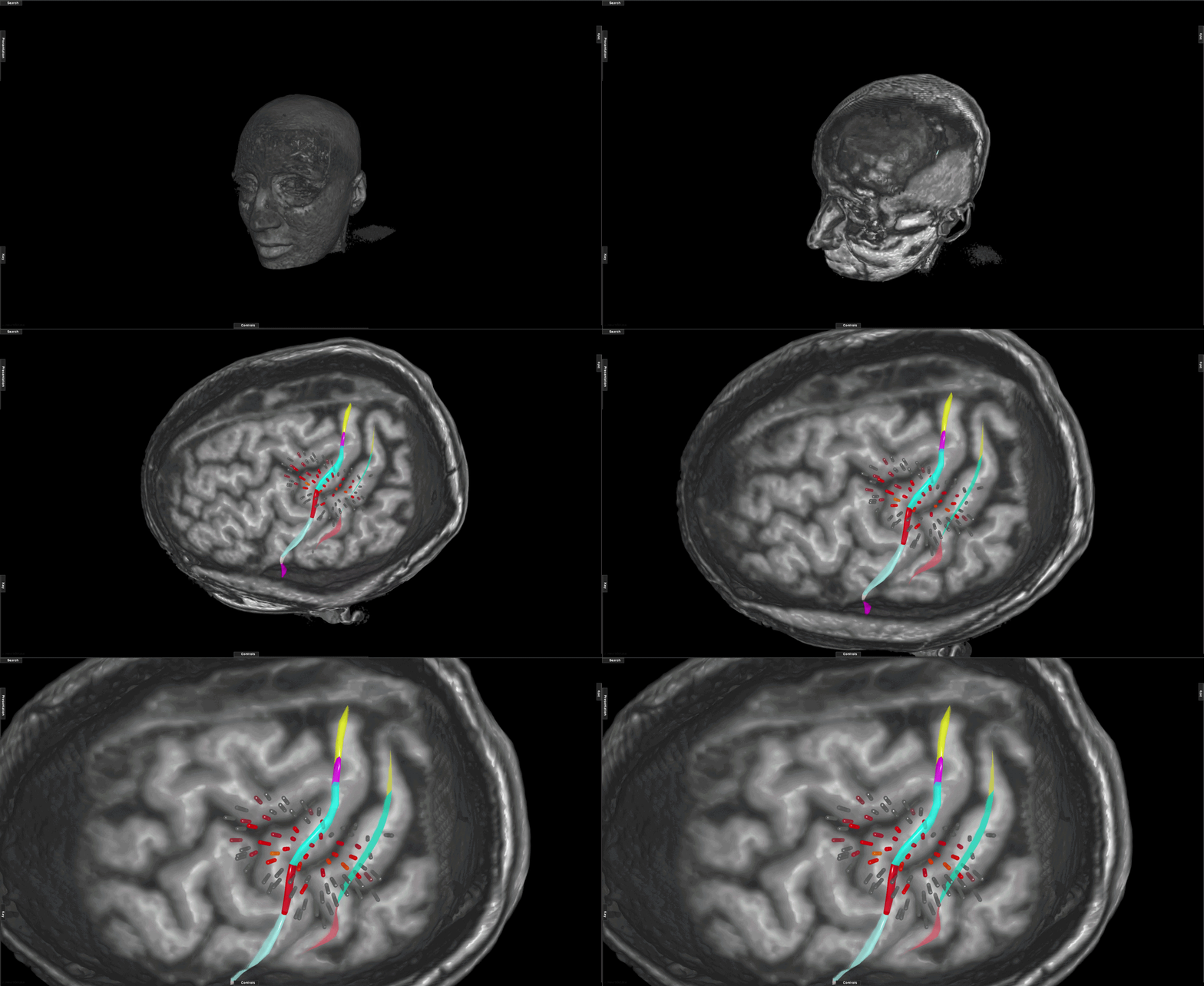
For drug-resistant epilepsy workflows that use stereoelectroencephalography, Neurospace can bring the implantation hypothesis, multimodal imaging, electrode trajectories, sampled regions, and intracranial findings into one shared spatial case. sEEG is treated as a diagnostic localization workflow—not simply another form of DBS.
- MRI, PET, SPECT, fMRI, DTI and other available case data in one context
- Multi-electrode stereotactic trajectory planning and visualization
- Vascular, anatomical and trajectory proximity review
- Seizure-onset and propagation network organization for multidisciplinary review
- AI-assisted correlation of imaging, anatomy, electrode locations and clinical findings
The platform supports case synthesis and planning context; seizure localization and treatment decisions remain specialist interpretations.
SEE THE LEAD.
UNDERSTAND ITS CONTEXT.
The existing Update 44 Functional Neurosurgery visualization is carried into this dedicated Space as a core DBS product interface. Here it becomes part of a broader workflow for relating the planned target and reconstructed lead position to deep anatomy, functional territory, selected pathways, contact locations, and the original planning hypothesis.
POST-PLAN INTELLIGENCE
POSITION IS ONLY MEANINGFUL IN CONTEXT.
Neurospace can preserve the relationship between the planned target, the reconstructed lead or electrode position, and the patient-specific anatomy used to create the plan.
This creates a common visual language for surgical review, multidisciplinary discussion and downstream analysis.
- Pre- and post-procedure image registration context
- Electrode-to-pathway relationship visualization
- Contact and proximity review
- Structured case documentation support
SEE AHEAD OF THE INCISION.
Functional procedures increasingly depend on understanding networks rather than coordinates alone. Neurospace Intelligence Layer can organize tractography and patient-specific spatial data around a candidate plan so the surgeon can compare how alternative targets or trajectories relate to selected pathways.
The goal is not to claim certainty where the data cannot provide it. The goal is to make relationships, assumptions and proximity visible so the clinical team can evaluate them explicitly.
MOVE FROM SLICES
TO SPATIAL UNDERSTANDING.
When spatial computing adds clinical value, Neurospace can present the patient-specific model through Mixed Reality for planning, rehearsal, education and collaborative review. AR is treated as a clinical viewing mode—not decoration.
THE PLAN, IN THE CLINICIAN'S FIELD OF VIEW.
Through a device such as Microsoft HoloLens 2, a clinician can inspect selected targets, pathways and trajectories as spatial objects while remaining anchored to the same planning dataset used on the workstation.
Remote specialists can review the same case through Neurospace Teleportal, annotate spatially and discuss alternative approaches without turning the session into a generic video call.
Illustrative clinician POV interface. Spatial overlays are visible through the mixed-reality device; Neurospace does not imply autonomous intraoperative decision-making.
ADOPT NEUROSPACE.
KEEP THE SYSTEMS YOU TRUST.
Functional Neurosurgery Space is positioned as an intelligence and visualization layer that can work with existing hospital infrastructure. It should help unify the case—not require institutions to discard established imaging, navigation or information systems.
PACS + DICOM
Ingest and organize available imaging through familiar clinical data standards and infrastructure.
MULTIMODAL IMAGING
Bring MRI, CT, diffusion and available functional datasets into a shared patient-specific coordinate space.
NEURONAVIGATION
Prepare reviewed planning information for connected surgical navigation workflows where compatible integrations are available.
TELEPORTAL
Extend the same case context to remote review, expert collaboration, training and multidisciplinary planning.
BUILT AROUND THE QUESTIONS FUNCTIONAL TEAMS ACTUALLY ASK.
Neurospace is framed around specialist workflow support rather than autonomous indications or regulatory claims. The same patient-specific environment can organize very different functional neurosurgical cases while preserving the distinction between diagnostic mapping, therapeutic stimulation, stereotactic intervention, and multidisciplinary interpretation.
DBS TARGET PLANNING
Patient-specific target, trajectory, anatomy, connectivity and risk review for deep brain stimulation planning.
EPILEPSY + sEEG PLANNING
Organize the implantation hypothesis, sampled regions, stereotactic electrode trajectories and multimodal imaging into one spatial case.
SEIZURE-NETWORK REVIEW
Relate intracranial electrode locations and clinical findings to anatomy and network context for multidisciplinary epilepsy review.
FUNCTIONAL MAPPING
Combine available fMRI, TMS, DTI, tractography and other functional data with structural anatomy for patient-specific interpretation.
LEAD LOCALIZATION + REVIEW
Relate post-procedure DBS lead position to the original target model, contacts, anatomy and selected pathways.
MULTIDISCIPLINARY CASE PLANNING
Create one shared visual context for neurosurgery, neurology, epileptology, neuroradiology, engineering and remote expertise when appropriate.
EVERY MILLIMETER
DESERVES CONTEXT.
Bring anatomy, function, connectivity, artificial insight and surgical experience into one patient-specific planning environment.